Reconstructive Surgery
Head, neck and jaw reconstruction
Tissue loss after removal of head and neck cancers is one of the most demanding areas of reconstruction in the body. Here the aim is not only appearance but the restoration of speech, chewing and swallowing.
Written and medically reviewed by: Doç. Dr. Tahsin Oğuz Acartürk — Plastic, Reconstructive & Aesthetic Surgery; University of Pittsburgh. Last updated:
Why these reconstructions are different
The head and neck contain many functions within a small volume: speech, chewing, swallowing, breathing and facial expression. Removing cancer with safe margins usually leaves a defect involving bone, muscle, mucosa and skin at the same time.
Repairing that loss is markedly harder than microsurgical reconstruction elsewhere in the body. The three-dimensional geometry of the region is complex, the transferred tissue must satisfy both form and function, and surgery often takes 10–12 hours — longer still in cases involving the skull base.
These operations are not the work of a single surgeon. They are planned together with ENT, surgical oncology, radiation oncology, dentistry, and speech and swallowing therapy teams. In centres unable to work across disciplines, the result is inevitably limited.
Tissue transfers used
Fibula free flap
The fibula in the lower leg is taken with its vessels and overlying skin paddle. It is shaped and plated to the contour of the jaw and connected to the neck vessels. Because it provides bone of a quality that can carry dental implants, it is the standard for reconstruction of the lower jaw.
Radial forearm flap
Its thin, pliable structure makes it the choice for reconstruction of the oral mucosa and tongue, preserving the mobility needed for speech and swallowing.
Anterolateral thigh flap
Used for large soft tissue defects. Its thickness can be adjusted and the donor scar is concealed on the thigh.
Scapula and latissimus dorsi flaps
Used in complex cases requiring bone and large-volume muscle together.
- Vessels of the transferred tissue are connected under the microscope at roughly 1–2 mm in diameter
- Tissue perfusion is monitored closely for the first 72 hours
- Secondary revision and dental rehabilitation are planned where needed
Surgery and recovery
- STEP 1 — JOINT PLANNINGThe area to be removed and the resulting defect are determined in advance with the oncology team; the donor site is selected.
- STEP 2 — SURGERYTumour removal and reconstruction are performed in the same session, with two teams working simultaneously.
- STEP 3 — FIRST 72 HOURSRegular monitoring of tissue perfusion in intensive care. This period is decisive for the outcome.
- STEP 4 — FIRST 6 WEEKSWound healing, nutritional support and the start of speech and swallowing therapy.
- STEP 5 — 3–12 MONTHSSecondary revisions where needed, dental implants and care of irradiated tissue.
Healing is slower and the risk of complications higher in tissue that has received radiotherapy. In such cases, fat tissue and stem-cell assisted treatments may be used as an adjunct to improve tissue quality.
Frequently asked questions
How long do these operations take?
On average 10–12 hours. In complex cases involving the skull base they can exceed 24 hours. The duration depends on the size of the defect and how many teams work simultaneously.
What happens if the transferred tissue fails?
The risk of flap loss in microsurgery is around 5% worldwide. When recognised early, the tissue can be salvaged by urgent vascular revision, which is why monitoring in the first 72 hours is critical.
Will my speech and swallowing return to normal?
That is the aim, but the outcome depends on the amount and site of tissue removed. Speech and swallowing therapy is an integral part of the process; regaining function may take months.
Can I have teeth fitted to my jaw?
Dental implants can be placed in a lower jaw reconstructed with a fibula flap. Timing is determined by bone consolidation and radiotherapy status.
Will taking bone from my leg affect my walking?
The fibula is not the main weight-bearing bone. Walking function is preserved after its removal; supported walking and physiotherapy are needed in the first weeks.
Doç. Dr. Tahsin Oğuz Acartürk
Plastic, Reconstructive & Aesthetic Surgery · Oral & Maxillofacial Surgery · University of Pittsburgh
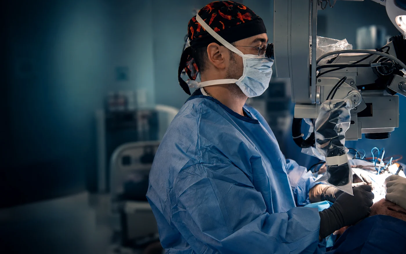
Experience in this field: Head and neck reconstruction lies at the centre of Assoc. Prof. Dr. Acarturk's academic career. In 2011 he was appointed Director of Head & Neck Reconstruction at the Department of Plastic Surgery, University of Pittsburgh. During that post he performed more than 300 microsurgical head and neck operations, achieving a rate of flap survival not previously reached in that department.
- Former Director of Head & Neck Reconstruction, University of Pittsburgh
- 600+ microsurgical head and neck cases
- Subspecialty in Oral & Maxillofacial Surgery — Turkish Ministry of Health
Assessment and second opinion
Send us your current diagnosis, the surgery planned or already performed, your radiotherapy history and any imaging. We will get back to you to coordinate with your oncology team.