Microsurgery Programme
Stages of lymphedema
Stage is the single most important piece of information determining the treatment option in lymphedema. The same swelling requires a completely different treatment at different stages. This page explains what the stages mean and what is possible in each.
Written and medically reviewed by: Doç. Dr. Tahsin Oğuz Acartürk — Plastic, Reconstructive & Aesthetic Surgery; University of Pittsburgh. Last updated:
The ISL staging system
The classification of the International Society of Lymphology (ISL) is the framework accepted worldwide. The stages describe the condition of the tissue and its reversibility.
| Stage | Clinical findings | Tissue state |
|---|---|---|
| Stage 0 (latent) | No visible swelling; the patient may feel heaviness | Transport capacity reduced, tissue normal |
| Stage 1 | Swelling increases through the day, partly resolves on elevation; pits on pressure | Fluid accumulation predominates, reversible |
| Stage 2 | Does not resolve on elevation; pitting gradually diminishes | Fibrosis and fat deposition have begun |
| Stage 3 (elephantiasis) | Marked increase in volume, thickened skin, wart-like changes | Advanced fibrosis, permanent tissue change |
Stage 0 is the most valuable stage. There is no visible swelling but a reduction in transport capacity is detected on imaging. In a patient caught at this stage, preventive and least invasive options are available. This is why regular follow-up matters in patients at risk.
Which treatment at which stage?
Stage directly determines which surgical option is applicable. The basic logic is this: if the lymphatic channels still work, redirect the flow; if they do not, bring in new tissue or reduce the volume.
| Stage | Priority approach |
|---|---|
| Stage 0 | Follow-up, skin care, risk reduction; preventive LVA in selected patients |
| Stage 1 | LVA — the least invasive and most effective option |
| Stage 2 | LVA and/or vascularised lymph node transfer |
| Stage 3 | Lymph node transfer + reduction surgery (liposuction, excision) |
This table is a framework, not a prescription. The final decision is made together with whether working lymphatic channels are visible on imaging, the duration of the swelling, the history of infection and the patient's general condition. Two patients at the same stage may be offered different operations.
How is the stage determined?
Staging is not done by looking alone; clinical examination and imaging are used together.
Clinical assessment
- Circumference measurements — compared with the opposite limb
- Pitting test — fluid or fibrosis
- Stemmer's sign
- Skin quality and thickness
- History and frequency of infection
Imaging
- ICG lymphography: a real-time map of working lymphatic channels. Decisive in planning LVA.
- Lymphoscintigraphy: assessment of lymphatic flow and nodal involvement
- MR lymphangiography: detailed anatomical imaging; in advanced and complex cases
Clinical stage and imaging findings do not always coincide. A patient who appears clinically stage 2 may still have working channels on ICG — which raises the possibility of LVA. For this reason a surgical decision should not be made without imaging.
Movement between stages, and time
Lymphedema is a progressive disease, but the rate of progression varies greatly from person to person. Some patients remain at stage 1 for years, while in others the picture advances rapidly.
Factors that accelerate progression
- Recurrent cellulitis attacks — the strongest accelerator
- Weight gain
- Irregular use of compression garments
- Time passed without treatment
- Additional trauma or surgery
Factors that slow progression
- Regular use of compression garments
- Skin care and infection prevention
- Weight control
- Regular movement and exercise
- Early surgical intervention
Can a stage be reversed? In the early stage — while fluid accumulation predominates — marked regression is possible with treatment. Once fibrosis is established, the tissue change is permanent; at this stage the aim is to reduce volume and halt progression.
Videos
Assoc. Prof. Acartürk Explains
Tap a cover to play the video right here. Videos are in Turkish.
 Lenfödemde cerrahi ne zaman: erken mi, geç mi?
 Lenfödemde tedavi seçenekleri
Frequently asked questions
How many stages of lymphedema are there?
Four, according to the International Society of Lymphology (ISL) classification: stage 0 (latent), stage 1, stage 2 and stage 3 (elephantiasis). The stages describe the condition of the tissue and its reversibility.
What does stage 0 mean?
There is no visible swelling, but lymphatic transport capacity is reduced and this is detected on imaging. The patient may feel heaviness or tightness. It is the stage at which treatment options are widest.
At which stage is surgery performed?
At stages 1 and 2, LVA comes to the fore if the lymphatic channels still work. At stages 2–3, vascularised lymph node transfer and reduction surgery are planned. The decision is made together with imaging findings.
Can a stage be reversed?
In the early stage, while fluid accumulation predominates, marked regression is possible with treatment. Once fibrosis is established the tissue change is permanent; at this stage the aim is to reduce volume and halt progression.
What happens if my stage advances?
Treatment options narrow. Whereas LVA, the least invasive method, is possible in the early stage, more extensive surgery becomes necessary in advanced stages. The risk of infection and loss of function also increase.
If it does not pit, is the stage more advanced?
Usually yes. A pit remains in the early stage; as the tissue hardens with fibrosis no pit remains. This is an important examination finding indicating that the picture has advanced.
Doç. Dr. Tahsin Oğuz Acartürk
Plastic, Reconstructive & Aesthetic Surgery · Oral & Maxillofacial Surgery · University of Pittsburgh
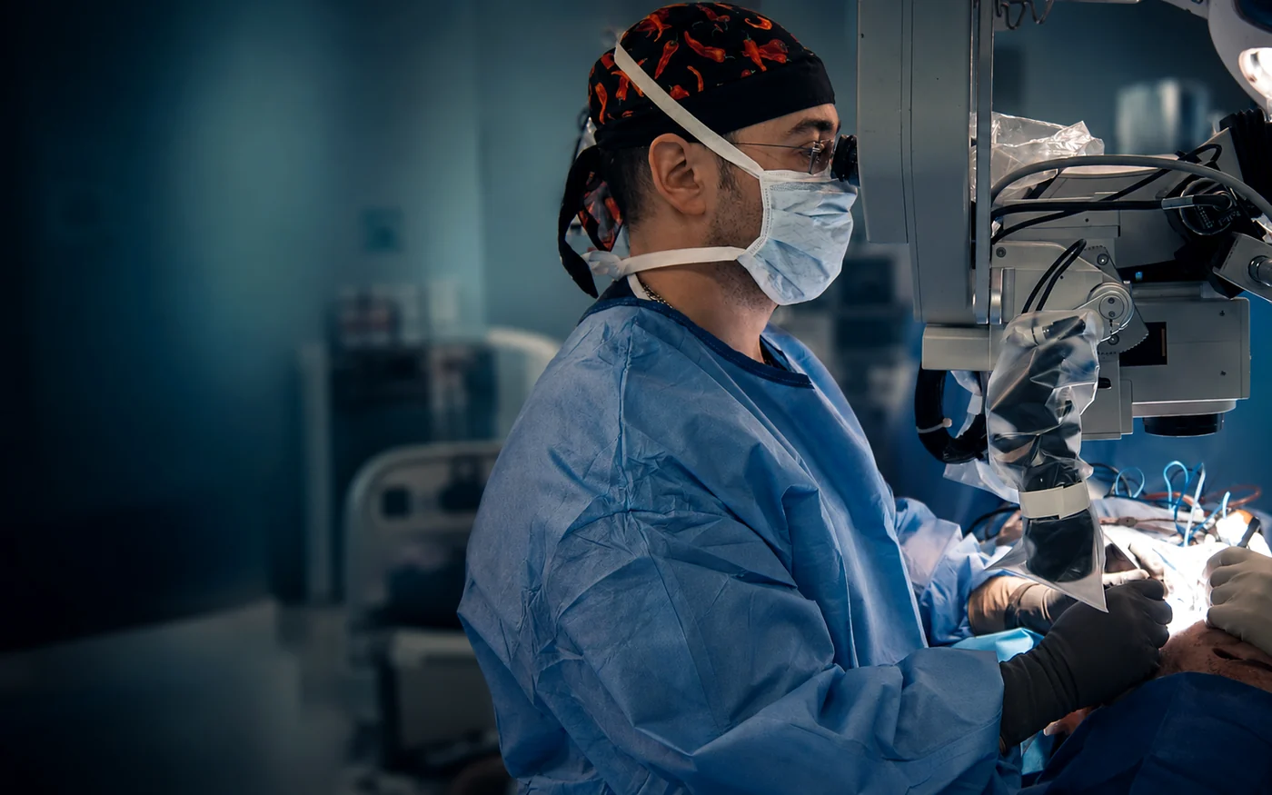
Experience in this field: Staging is the framework that determines treatment but is not sufficient on its own — it must be interpreted together with imaging findings. As a microsurgeon who participates in the work of the European lymphatic microsurgery group and has presented at the World Congress of Lymphology, Assoc. Prof. Dr. Acarturk assesses these two sets of data together.
- Participation in the European lymphatic microsurgery group
- Presentation at the World Congress of Lymphology
- 600+ microsurgical cases at the University of Pittsburgh
Let us determine your stage together
Tell us how long the swelling has lasted, whether it resolves on elevation, your history of cellulitis and any imaging results you have; attach a photograph if possible.